Developments in the treatment of congenital limb deficiencies and deformities have accelerated, especially with the invention of the Ilizarov technique in the middle of the last century. Thanks to this technique, bone regeneration is provided in the human body and congenital limb deficiencies are treated with these bones. However, this method discovered by mankind cannot create a bone tissue in the shape and naturalness of the bone developing in the womb. Therefore, these bones and tissues are shaped as a result of long efforts and functional functioning is provided with complex techniques.
Congenital limb deficiencies appear clinically in different forms. It is in the form of clubhand in the arms, ‘Tibial Hemimelia’, ‘Fibular Hemimelia’ in the legs, lack of fingers in the hands and feet, cleft foot, cleft hand. These disorders can be seen together or in isolation. For example, in the ‘Tibial Hemimelia’ disorder, the ‘tibia’ bone, which is popularly known as the shin bone in the body, is deficient in a difference between a small amount of lack of development and complete absence. The greater the bone deficiency, the lower the chance of achieving a functional leg with treatment. When these babies are born, the distortion of their feet is visible. Early diagnosis can be made by careful examination of the feet during the first examination.
In ‘Fibular Hemimelia’, which is another disorder, there is a deficiency in the ‘fibula’, the cane bone next to the shin bone. As in the tibia, there may be varying degrees of defects, ranging from complete absence of bone to a small amount of bone deficiency. As the amount of deficiency increases, treatment becomes more difficult. Just like in the tibia, fibula deficiencies also cause a difference in the feet at birth. Sometimes it can even be confused with clubfoot, which can be treated more easily. In clubfoot, there is no deficiency in the bones.
Sometimes deficiencies can be seen where the thigh bone meets the hip joint. This type of deficiency, known as ‘PFFD’ (Proximal focal femoral deficiency), ‘PFD’ (Proximal femoral deficiency) or ‘Congenital Femoral Insufficiency’, can cause the hip joint to not form at all and requires both bone shaping and joint formation surgery. Since the body’s own healing potential is used in bone formation, repeated surgeries and lifelong planning are required in the treatment of almost all congenital diseases.
Regardless of the bone deficiency, the treatment process is long and requires repeated surgeries. No matter how willing the family is to undergo treatment, some defects cannot be corrected, even in rare cases. In these cases, regardless of the centre in the world, amputation (removal of the unusable part of the leg) results. This treatment, which was more preferred in the past years, is now very rarely applied today thanks to the successful results and advanced techniques in reshaping surgery.
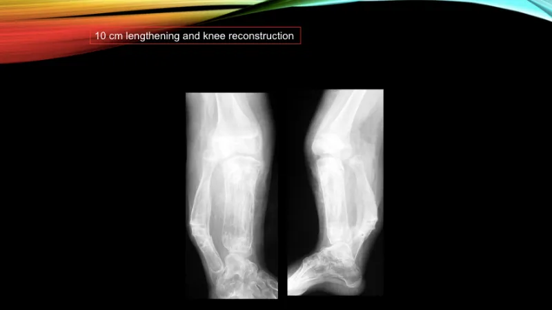
In congenital limb deficiencies, a detailed history is taken from the moment of the first encounter. The condition of the bones and joints is evaluated. After the necessary radiological studies, it is decided whether reshaping and limb salvage can be performed. If rescue and reshaping is to be performed, the number of operations to be performed and the most appropriate age intervals for the operations are determined. A lifelong treatment plan is explained to the family and presented in detail. After the family understands this long treatment period and declares their compliance and acceptance, the operations are started. In the treatments, internally or externally implanted fixation devices, specially made walking orthoses, compensated elevators, physical therapy and rehabilitation programmes are applied. At the end of all these treatments, a functional, complete limb shaping that enables walking is performed.
Although all these procedures are performed with absolute care, experience and in accordance with basic principles, undesirable situations called complications may occur during surgery or during treatment. The most common complications are vascular nerve damage during osteotomy, bone and nail bottom infection, non-union or late union at the osteotomy site, joint movement limitation, subsequent fracture at the osteotomy site, etc. However, one of the important advantages of this method is that the complications that may occur are eliminated within the treatment period and with the same device. On the other hand, psychological and social support should be provided to both parents and children during treatment in order to ensure absolute compliance in paediatric patients.
In conclusion, the treatment of congenital limb deficiencies is planned according to the anatomy and deficiency of each patient, close follow-up and sequential surgeries are applied until growth is completed, family and patient compliance is at the forefront, rehabilitation programmes are needed, laborious and long treatments. It should not be forgotten that a devoted and experienced surgical team is needed for these treatments which are successfully applied in our country.
TIBIAL HEMIMELI
PRELIMINARY REPORT ON AMPUTATION VERSUS RECONSTRUCTION IN TREATMENT OF TIBIAL HEMIMELIA ACTA ORTHOP TRAUMATOL TURC 2015;49(6):627–633 DOI: 10.3944/AOTT.2015.15.0005
HALIL İBRAHIM BALCI, YAVUZ SAĞLAM, FUAT BİLGİLİ, CENGIZ ŞEN, MEHMET KOCAOĞLU, LEVENT ERALP
ISTANBUL UNIVERSITY ISTANBUL FACULTY OF MEDICINE, DEPARTMENT OF ORTHOPAEDICS AND TRAUMATOLOGY, İSTANBUL, TURKEY
OBJECTIVE: TIBIAL HEMIMELIA IS A RARE DISORDER CHARACTERIZED BY THE ABSENCE OR HYPOPLASIA OF THE TIBIA WITH ASSOCIATED RIGIDITY. THE AIM OF THIS STUDY WAS TO RETROSPECTIVELY EVALUATE THE AFFECTIVITY OF RECONSTRUCTIVE SURGERIES INCLUDING CENTRALIZATION OF THE KNEE-ANKLE JOINTS AND LENGTHENING WITH ILIZAROV PRINCIPLES, AS WELL AS PHYSICAL AND FUNCTIONAL RESULTS OF AMPUTATION AND RECONSTRUCTION. METHODS: THIS IS AN IRB-APPROVED RETROSPECTIVE REVIEW OF ALL PATIENTS DIAGNOSED WITH TIBIAL HEMIMELIA WHO REQUIRED SURGERY AT A SINGLE INSTITUTION BETWEEN 1998 AND 2011. CHARTS WERE ANALYZED FOR CLINICAL AND RADIOGRAPHICAL FINDINGS. AT FINAL FOLLOW-UP, PATIENTS UNDERWENT PHYSICAL AND RADIOGRAPHIC EXAMINATION. PATIENTS AND THEIR PARENTS WERE ASKED TO COMPLETE THE SF-10™ HEALTH SURVEY (QUALITYMETRIC INC., LINCOLN, RI, USA). RESULTS: TWENTY-ONE PATIENTS (12 MALE, 9 FEMALE) WITH 30 AFFECTED EXTREMITIES WERE INCLUDED. MEAN AGE WAS 4.8±3.1 YEARS AT INITIAL SURGERY. KNEE LEVEL DISARTICULATION WAS PERFORMED IN 6 EXTREMITIES OF 4 PATIENTS. ONE PATIENT WITH TYPE III UNDERWENT TRANSTIBIAL AMPUTATION. MEAN NUMBER OF SURGERIES FOR EACH PATIENT WAS 6.4±3.3, AND MEAN DURATION OF EXTERNAL FIXATOR AND CASTING WAS 17±6 MONTHS. MEAN LENGTHENING WAS 4.9±1.3 CM, AND MEAN LIMB LENGTH DISCREPANCY WAS 3.1±1.7 CM AT 5.8±3.7 YEARS AT FOLLOW-UP. SF-10™ SCORES WERE SIMILAR IN DISARTICULATED AND RECONSTRUCTED PATIENTS (P=0.63). ALL SCORES WERE SIGNIFICANTLY HIGHER WHEN DISARTICULATION WAS PERFORMED IN CASES OF KNEE INSTABILITY (P<0.01). CONCLUSION: WHEN STABILITY OF THE KNEE JOINT IS PRESENT, TREATMENT MODALITY SHOULD BE CHOSEN ACCORDING TO THE EXISTENCE OF THE PROXIMAL TIBIA. AMPUTATION SHOULD BE PREFERRED IN CASES OF KNEE JOINT INSTABILITY. KEYWORDS: AMPUTATION; EXTERNAL FIXATION; KNEE CENTRALIZATION; RECONSTRUCTION; SF-10™; TIBIAL HEMIMELIA. LEVEL OF EVIDENCE: LEVEL III THERAPEUTIC STUDY